Sheryl Shook
Heart Dissection
Learning Objectives
- Demonstrate proficient dissection skills.
- Identify external anatomical structures of the heart.
- Identify internal anatomical structures of the heart.
1. Instruments
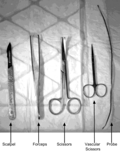


Images by Karolinska Institutet / CC BY 4.0
2. Surface anatomy of the heart
Begin by orienting the heart (Figure 5.3). Take note of its size and shape. Specimens may or may not have been perfused with a preservative that will cause a different heart color than shown. Identify the following landmarks:
- Apex
- Auricles
- Demarcation between the right and the left ventricle (interventricular septum)
- Entrances of superior vena cava and inferior vena cava
- Coronary blood vessels
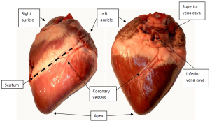
Image by Karolinska Institutet / CC BY 4.0
The heart commonly has some adipose tissue on the outer surface. You also may find cuts that have been made when the heart was removed from the chest cavity.
The auricles are appendages to the atria. Their function is not entirely known. It has been speculated that they function as reservoirs for blood, which may be mobilized in times of increased physiological need.
3. Aorta and the pulmonary arteries
Several vessels extend from the superior (topmost) side of the heart. It may be necessary to remove excessive tissue to visualize structures and for ease of dissection. Identify the aorta and the pulmonary artery and then cut them to make the valves visible (Figure 5.4). The aortic and pulmonary valves are each made up of three leaflets. Inspect the valves and palpate them (feel them with a finger).
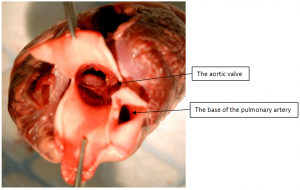
Image by Karolinska Institutet / CC BY 4.0
Try pouring a little water on the valve and see if the valve closes.
Thought question:
Why do the arteries leading blood from the heart have thicker walls than the veins leading blood back to the heart?
4. Coronary arteries
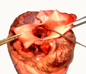
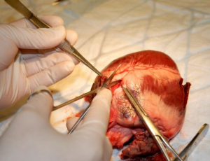
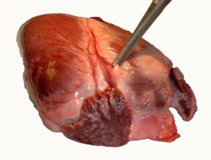
The two main coronary arteries branch off from the aorta just above the aortic valve. Start by probing the coronary artery entrance (Figure 5.5). By gripping the heart with the forceps next to the entrance of the artery, you may straighten out the closest part of the artery, which makes it easier to cut. Cut the coronary arteries starting from the aorta and proceeding along one or more branches as far as their size permit (Figure 5.6). Use the forceps to visualize the opened the artery (Figure 5.7).
Images by Karolinska Institutet / CC BY 4.0
Thought question:
In angina or myocardial infarction, the myocardium (heart muscle) receives insufficient blood flow. The effect of diminished flow in one coronary artery depends on, among other things, whether other arteries can compensate for the decreased flow. The anatomy of the coronary vessels may vary greatly between individuals. How much of the heart appears to receive its blood supply from only one single artery?
5. The right atrium
Identify the openings for the superior and inferior vena cavae (Figure 5.8). Cut open the right atrium along the path between the vena cavae openings and continue into the auricle (Figure 5.9). Remove any coagulated blood that may be found here. Take note of the smoothness of the endocardium, the tissue lining the inside of the heart. Also notice the pectinate muscles.
During fetal life, when lungs are not used for gas exchange, there is an opening—foramen ovale. This opening normally closes at birth, leaving a shallow pit—fossa ovalis—in the myocardium. Sometimes, a small opening may persist, and is then termed a patent foramen ovale. Find the fossa ovalis and examine with a probe whether any opening remains (Figure 5.10).
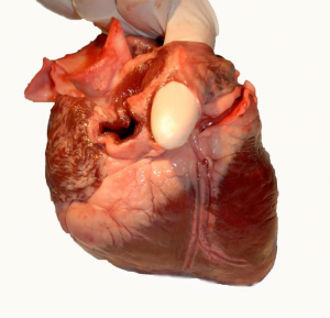
Image by Karolinska Institutet / CC BY 4.0
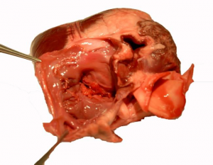
Image by Karolinska Institutet / CC BY 4.0
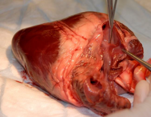
Image by Karolinska Institutet / CC BY 4.0
Thought question.
Deep venous thromboses are not uncommon. They may give rise to a thromboembolism, when part of a blood clot comes off, and is transported by the blood to some other part of the body thus blocking the blood flow. How would a patent (open) foramen ovale affect the range of possible outcomes?
6. The right ventricle
Cut open the right ventricle through the opening from the right atrium, along the right side of the heart (Figure 5.11). You will then cut through the tricuspid valve. Open the right ventricle and take note of the valve (Figure 5.12). This valve has three cusps, which are attached to papillary muscles extending from the inner wall of the ventricle. Observe and palpate the valve (Figure 5.13). Proceed to examine the muscular wall of the ventricle and take note of the trabeculae carneae (beam-like structures). Finally, identify the pulmonary valve and probe it from the inside of the ventricle.
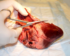
Image by Karolinska Institutet / CC BY 4.0
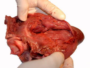
Image by Karolinska Institutet / CC BY 4.0
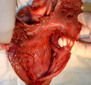
Image by Karolinska Institutet / CC BY 4.0
Thought question:
Can you trace the blood flow out of the right ventricle? What organ is the first to receive this blood?
7. Left atrium and ventricle
Cut open the left atrium and ventricle through the entrance for the pulmonary vein into the left atrium (Figure 5.14). Cut along the left side of the heart. The valve between the left atrium and ventricle has two cusps and is termed the bicuspid or mitral valve (miter = type of hat that tapers to a point). Examine the endocardium and the mitral valve (Figure 5.15). Identify the entrance to the aorta and probe it from the inside of the left ventricle.
Compare the right and left ventricles with regard to their respective sizes and the thickness of the myocardium. Finally, palpate the muscle wall between the right and left ventricles (interventricular septum). Here run the right and left bundle branches, part of the electrical conduction system of the heart.
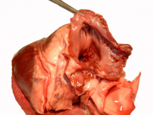
Image by Karolinska Institutet / CC BY 4.0
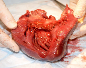
Image by Karolinska Institutet / CC BY 4.0
Thought question:
Why is the myocardium of the left ventricle thicker than that of the right ventricle?
“Heart Dissection” is MODIFIED from:
- Dissection manual for porcine heart by Karolinska Institutet / CC BY 4.0
- Text: Gustav Nilsonne
- Photos: Lotta Arborelius
